


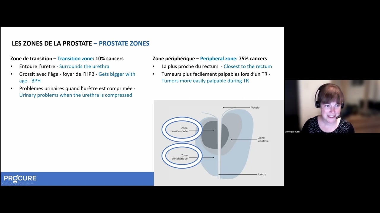
Abnormalities detected during a digital rectal exam and a high PSA level often lead to a prostate biopsy. This procedure consists of taking small tissue samples of your prostate in order for the pathologist to examine them under a microscope to determine if they are cancerous or not. If cancer is detected, the pathologist will use the same samples to calculate your grade (or Gleason Grade Score).
A prostate biopsy is usually performed with the help of a transrectal ultrasound (TRUS biopsy). The images taken with the ultrasound help guide a fine needle to the areas selected for sampling. The spring-loaded needle is attached to the ultrasound probe and enters the prostate through the rectum. Usually between 6 – 12 (sometimes more) prostatic tissue samples are obtained and the entire procedure lasts about 10 minutes. A local anesthetic can be used to numb the area and reduce any pain.
Even though they are very useful, the digital rectal exam and various PSA tests are not sufficient to diagnose prostate cancer.
In the days following a transrectal biopsy, blood may be visible in your excrement. It is possible that you notice reddish or brownish blood in your urine and sperm for several weeks. Light bleeding is normal. However, if it becomes more severe, takes longer to clear up, or if there is a lot of thick-looking, clotted blood in your urine or stools, you should contact your doctor or go to the nearest emergency department.
In the days following your procedure, you may experience pain or discomfort in the biopsy area. Pain medication can be prescribed if feel that you need them.
There is a small risk of infection after a biopsy – bacteria on the biopsy needle can get into the prostate. To reduce the risks of infection, antibiotics may be prescribed before and after the procedure. Fever, chills, pain, or burning during urination are signs of infection that can happen even if you are taking antibiotics. If you have these symptoms, contact your doctor or go to the nearest emergency department.
Urine retention is the inability to pee despite having a full bladder. A small percentage of men suffer this side effect following a prostate biopsy. If you suffer from urine retention, speak with your doctor or go to the nearest emergency department.
It takes about two weeks to get the biopsy results because the pathologist needs to examine the samples under a microscope before giving his report.
If the pathologist finds cancer in the samples, he will assess the aggressiveness of the cancer by giving it a Gleason grade. You will discuss possible treatments with your urologist who may recommend other complementary tests in order to determine how far your cancer has progressed.
The fact that the pathologist did not find any cancer in your samples can be reassuring. However, it is possible that the biopsy needle simply missed the cancerous areas and you do in fact have cancer. If your urologist suspects that you have cancer, he may have you do a second transrectal biopsy or another type of prostate biopsy. Otherwise, regular prostate checkups, including the PSA test, digital rectal exam (DRE), and magnetic resonance imaging (MRI), may be the preferred route.
While the transrectal prostate biopsy is the most common, there are other types of biopsies that your urologist may perform.
Done under local or general anesthesia, this biopsy takes prostate samples by inserting a needle through the skin of the perineum (the region between the scrotum and rectum). As this procedure allows a larger area of tissue to be examined than a transrectal biopsy, your urologist may order one if they think you have cancer despite a negative transrectal biopsy.
Done under local or general anesthesia, this biopsy goes through the urethra (the tube that runs from the penis to the bladder) to collect the prostate samples. To do this, the urologist inserts a thin tube with a camera attached (a cystoscope) into the penis. A surgical tube is then passed through the cystoscope to collect the samples.
If the biopsy confirms the presence of a tumour, additional examinations may be required to determine whether the cancerous cells have spread elsewhere in the body. These are generally undergone by men presenting serious signs of the disease, such as an extensive induration or lump in the prostate, elevated PSA levels or biopsy results indicating the presence of an aggressive cancer. Some of these tests will help the characterization and staging of your cancer.
This test, done by taking a blood sample, is not used for staging; it is used to evaluate your overall health by measuring the number and quality of your blood (red blood cells, white blood cells, and platelets). The complete blood count, among other things, checks for infection and anemia that can affect your treatment.
These tests measure the concentration of various chemical substances in the blood. These chemicals show how well your organs are functioning and can be used to detect damaged tissues or abnormalities. The following chemicals can help stage prostate cancer:
A bone scan is an imaging technique used to detect the presence of cancerous cells in the bone where prostate cancer most often spreads. Your urologist will usually order a bone scan if your alkaline phosphatase or calcium levels are elevated or if you have bone pain, or if your urologist has diagnosed you with a more aggressive prostate cancer.
A small amount of radioactive material (tracer) is first injected into your blood. You will need to wait two to three hours, the time it takes for the tracers to collect on your bone. During this time, you may need to drink a lot of liquids in preparation for the scan which will last 30 to 45 minutes. During the scan itself, you will lie on an examination table and a camera will take images of your body by moving slowly around you without touching you.
If the images show that the radioactive tracer is spread evenly over all of your bones, your results are normal. However, if prostate cancer has spread to the bones, we usually find it to be concentrated (darker areas) along the spine, ribs, or long bones.
Computed tomography, often called a CT scan, is also known as a computerized axial tomography (CAT) scan. A CT scan is a form of computed radiography (X-ray) that takes a 3-dimensional image of the internal organs in your body. As prostate cancer often spreads to the pelvic and abdominal nodes, your urologist may be particularly interested in knowing whether the cancer has spread to the smaller organs of your immune system. CT scans can also be used before a radiotherapy treatment to know the prostate’s exact dimensions.
For a CT scan, you will probably need to fast for a few hours (no solids or liquids). Before the CT scan, a non-radioactive colorant that helps to see the internal organs more clearly is injected. During the procedure, which lasts about 20 minutes, you will be lying still on a table that will slide slowly into the donut-shaped machine. A camera located on the inside of the machine’s ring will take a series of images of your entire body without ever touching you. You may be asked to hold your breath at times in order to take clearer images. Once completed, computer software will put these pictures together to form a three-dimensional image of the area of interest.
From the images taken, a urologist can see the size and shape of your prostate and determine, among other things, the efficiency of a given treatment. Enlarged lymph nodes could be an indication that they have metastasized and become cancerous. This may affect your course of treatment and other tests may be necessary.
Magnetic resonance imaging (MRI), often clearer and more defined than a CT scan, uses magnetic forces and radio-frequency waves rather than X-rays to take a 3-dimensional picture of your internal organs. An MRI lets you see if the prostate cancer has spread to other tissues like the bones, lymph nodes, or seminal vesicles. The MRI can also be used to plan your treatment, to evaluate the efficiency of a treatment, or to follow the cancer’s progression.
Before starting the procedure, which takes about an hour, you may be injected with a non-radioactive contrast substance in order to improve image quality. During the MRI, you will lie still on an examination table that slides into a donut- or tunnel-shaped machine. Inform your urologist if you are claustrophobic. Sometimes you may be asked to hold your breath in order to take clearer images. At the end, computer software will put these pictures together to form a three‑dimensional image of the area of interest.
From the images taken, a urologist can see the size and shape of your prostate and determine, among other things, the efficiency of a given treatment. The urologist will also be able to see if the cancer has metastasized out of the prostate which can impact your chosen treatment.
We invite you to consult our page Questions to ask to your doctor and your healthcare team regarding tests and diagnostic exams for prostate cancer. Asking questions will open up communication, provide information tailored to your situation, and reduce the stress associated with understanding the diagnosis of prostate cancer.

Learn more about the role of the urologist and the importance for a patient to gather adequate information after receiving a prostate cancer diagnosis.

A man with prostate cancer shares the challenges of his cancer experience.

You’ve been diagnosed with prostate cancer? Your role is as important as that of your medical team.

Recently diagnosed with cancer? Educate yourself to fully understand your situation.

After a prostate cancer diagnosis, questions about survival, cure rates, and quality of life can arise.

This webinar covers various tests and exams for diagnosing and monitoring prostate cancer.
This webinar clarifies biopsy reports, pathologists’ roles, and how results confirm or rule out prostate cancer.

After receiving a diagnosis of prostate cancer, you will undoubtedly have questions about your prognosis and survival and will want to know the chances of success with your treatment.

Do you have a significant family history of cancer? Is there a link between prostate cancer and a genetic mutation?

Prostate cancer often overwhelms patients. Finding the right words is crucial to comfort and support them.